|
DIARRHOEA Note for Pet Owners:
Topics on this Page: |
Description
The definition of diarrhoea is as follows:
Normal bowel movements are altered resulting in an increase in any one of the following:
- Frequency of defaecation.
- Fluid content in the stools causing a loose consistency. (NB The water content in normal faeces is 60-80%and 70-90% in loose stools).
- Volume of faeces produced - this is a combination of water content and undigestible (e.g. dietary fibre) or unabsorbed nutrients (as seen in malabsorption syndrome).
Appearance
The definition of diarrhoea does not cover the appearance of the stool - and it should be realised that not all forms of diarrhoea result in loose stools. The stool can still be formed, as it is with
Sometimes mucous will be seen, and sometimes fresh blood (see dysentery and haematochezia and melaena).
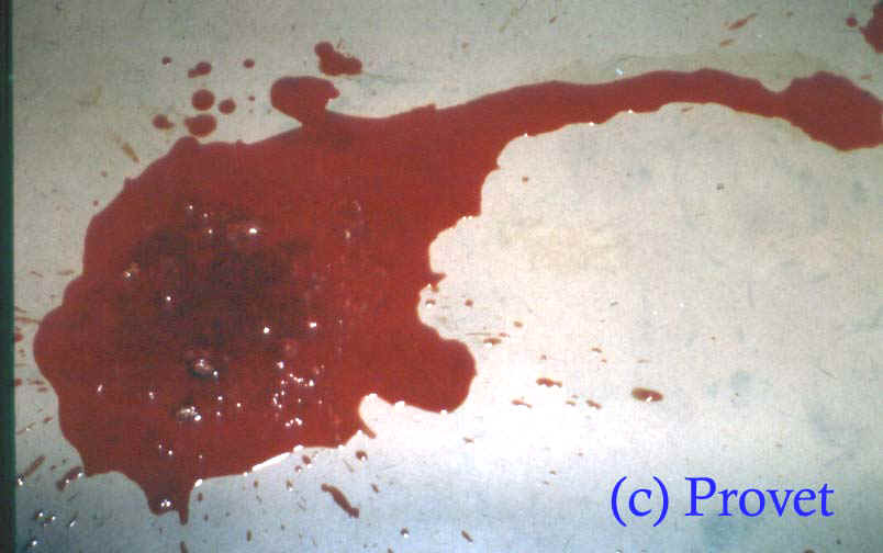
The photograph below is of dysentery
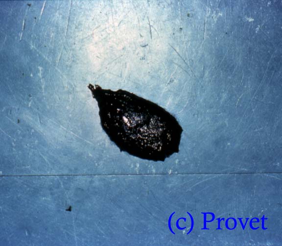
The photograph below is of black, tarry melaena
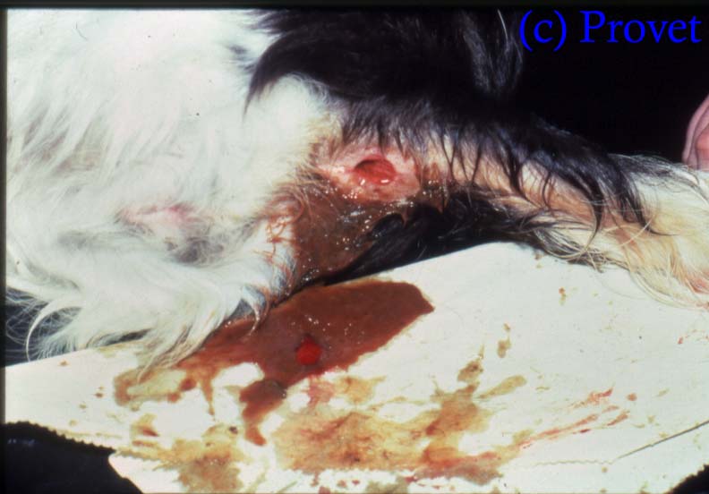
The photograph below is of haematochezia
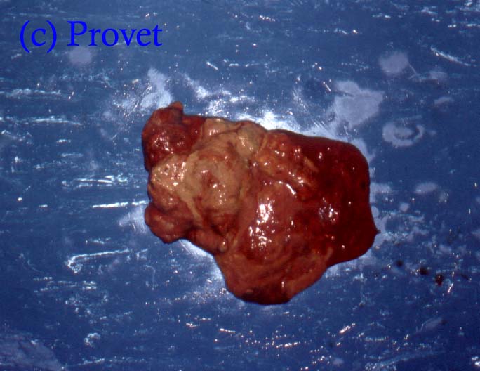
The picture below is of faeces, blood and mucus typical of colitis.
Sometimes blood can be present in a faecal stool without there being any obvious external change in appearance - this is called occult blood, and can be detected using sensitive laboratory tests.
The appearance of the diarrhoea can give a very good idea of the location of the gastrointestinal disorder:
| Appearance of faeces | Location of disorder |
| Normal colour. Increased volume. Large, bulky soft stool. | Small intestine |
| Very watery - no obvious blood | Small intestine |
| Very watery stool with fresh blood (dysentery) | Small intestine. Typical of canine parvovirus infection or Haemorrhagic gastroenteritis |
| Yellow, brown or green soft, bulky stool | Small intestine |
| Pale, greasy, putty-coloured, soft, fatty foul smelling stool | Steatorrhoea |
| Black, tarry stools. | Melaena |
| Very pale, white, crumbly stools | Lack of bile pigment |
| Small volume, but frequently passed stools | Colon |
| Formed faeces or watery faeces with mucus | Colon |
| Clear mucus or mucus | Colon |
| Watery faeces with fresh blood and mucus | Colon |
Cause
Diarrhoea is a very common clinical sign which can be associated with a whole variety of causes including:
| FUNCTIONAL CAUSE | REASON | UNDERLYING CAUSE |
| Osmotic movement of water into the faeces | Excessive food intake, overloading the system with retention in the intestine lumen. | Can be just excessive intake of one nutrient e.g. high fat content |
| Failure to digest nutrients properly resulting in retention in the lumen of the intestine | Exocrine pancreatic insufficiency, bacterial overgrowth, enzyme deficiency (e.g. lactase deficiency), bile salt deficiency | |
| Dietary intolerance | ||
| True dietary allergy (rare) | ||
| Diseases of the mucosal lining of the small intestine | Inflammation, neoplasia (cancer), gluten enteropathy (Irish Setters) | |
| Diseases of the wall of the intestine | Chronic enteritis, chronic cellular infiltration, lymphangiectasia, cancer (e.g. lymphosarcoma), canine sprue | |
| Secretory diarrhoea due excessive fluid secretion into the faeces | Secretory stimulants | Endotoxins produced by bacteria in the intestine |
| Hydroxylated bile acids in the intestine lumen | ||
| Inflammation of the intestine | Bacteria,Parasites, Viruses | |
| Increased permeability of the intestine wall | Lymphangiectasia , protein-losing enteropathy | |
| Motility disorders of the intestine | Reduced intestinal transit time | Low fibre diets, partial obstruction of the intestine |
| Rapid intestinal transit time | Many diseases of the intestine |
Diagnosis
The primary cause of a diarrhoea is not always possible to detect - particularly in acute diarrhoea when eating rubbish, a high fat snack or an unusual foodstuff might not have been noticed by the owners.
In chronic diarrhoea the appearance of the stool is very helpful in identifying the likely site of the problem, and the following tests can help with identifying an underlying cause:
| TEST | Exocrine Pancreatic Insufficiency | Proximal Small Intestine Disease * | Distal Small Intestine Disease * | Bacterial Overgrowth |
| TLI Test | Low | Normal | Normal | Normal or Increased |
| Cobalamin | Normal | Normal | Low | Low |
| Folate | Normal | Low | Normal | High |
| Bile acids | Normal | Normal | Low | Low |
| Faecal Fat | Increased | Normal | Increased | |
| Absorption Test (e.g. xylose) | Decreased | Decreased | Normal | Decreased |
Consequences
A patient can lose large amounts of water in diarrhoea and this causes dehydration which, in severe cases such as canine parvovirus disease, can be life-threatening.
Last updated : October 2013