RADIOGRAPHY OF THE CANINE LARYNX First broadcast on www.provet.co.uk |
This information is provided by Provet for educational purposes only.
You should seek the advice of your veterinarian if your pet is ill as only he or she can correctly advise on the diagnosis and recommend the treatment that is most appropriate for your pet.
The folds of the larynx overlapping the walls of adjacent structures creates a complex photographic image that makes interpretation of radiographs of this region quite difficult. Also, it is an anatomical region that clinicians infrequently radiograph.
Radiographic examination of the larynx is important in animals that have dysphagia - with apparent signs of pharyngeal retching, or regurgitation, or coughing following eating or drinking. It is also important when investigating suspect foreign bodies - or foreign body penetrations at the back of the throat, for suspect fractures of the hyoid bone or following other trauma to the region.
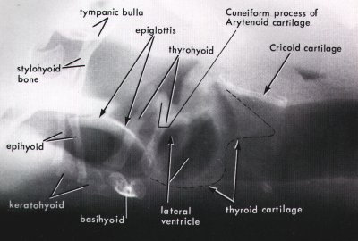
This labelled XRay helps to identify the major structures of a canine normal larynx and provides a useful reference with which to compare radiographs from clinical cases
Reproduced from O'Brien "Radiographic Diagnosis of Abdominal Disorders in the Dog and Cat" with the kind permission of Harcourt Brace Publishers (out of print)
This radiograph demonstrates the importance of maintaining a comprehensive library of radiology books or other reference plates with which clinical radiographs can be compared.
Last updated : October 2013