
|
HEART FAILURE
This information is provided by
Provet for educational purposes only.
You should seek the advice of
your veterinarian if your pet is ill as only he or she can correctly advise
on the diagnosis and recommend the treatment that is most appropriate for
your pet. Note for Pet Owners: Topics on this Page:
|
Description
Heart failure occurs when the heart fails to pump sufficient blood around the body to provide the oxygen and nutrients and to remove waste products that tissues need. This reduces the quality of life for the patient and can lead to reduced lifespan.
Congestive heart failure describes that form of heart failure when tissues become congested with fluid because of a failure in venous return to the heart - called right-sided heart failure.
Left-sided heart failure results in a failure to maintain sufficient output of blood from the heart (reduced cardiac output). Many animals have both left-sided and right-sided heart failure at the same time.
Cause
- Failure of the heart muscle
- Excessive circulating blood volume
- Excessive pressure on the heart
Breed Occurrence
There is no breed susceptibility to develop heart failure - though some breeds
are more susceptible to specific diseases which can lead to heart failure
developing than other breeds.
Signs
1. Right-sided heart failure (backward or congestive failure) causes the following signs :
|
Sign |
Technical term |
Underlying cause |
|
Cough - often after a period of rest (lying down) or during exercise |
Cough - sometimes paroxysmal |
|
|
Breathlessness |
|
Fluid accumulation (oedema) in the lung tissue |
|
Increased respiratory rate - panting Difficulty breathing |
Tachypnoea Dyspnoea |
Fluid accumulation (oedema) in the lung tissue (interstitial and/or alveolar) OR free fluid in the chest cavity (pleural effusion) - common in cats. |
|
Blue lips and other mucous membranes, abnormal chest noises, wheezing |
Cyanosis Noises - crackles, rhonchi |
Poor oxygenation of blood Fluid accumulation in the lung tissue (oedema in the alveoli and airway) |
|
Swollen jugular veins in the neck Swollen abdomen - enlarged liver or free fluid in the abdominal cavity |
Jugular distension
Hepatomegaly Ascites |
All due to right-sided heart failure. Fluid in the pericardial sac causing compressive pressure (tamponade) on the heart, leaking of the right atrioventricular valve (the tricuspid), dilated cardiomyopathy |
|
Heart enlargement - on X-ray |
Cardiomegaly |
Volume overload, fluid retention |
|
Abnormal heart sound (murmur) when examined with a stethoscope |
Gallop sound - due to third heart sound S3 |
Sound of blood filling the dilated ventricular chamber in the heart. |
|
Abnormal heart sound (murmur) |
Split second heart sound (S2) |
Delayed emptying of right ventricle resulting in delayed pulmonic valve closure. |
2. Left-sided heart failure (forward failure) causes the following signs
|
Signs |
Technical term |
Underlying cause |
|
Reluctance to exercise, or fatigue during exercise |
Exercise intolerance |
Poor oxygen supply to the muscles |
|
Pale lips and other visible mucous membranes |
Pallor |
Constriction of the blood vessels causing poor blood circulation to the area. Caused by increased sympathetic nervous system activity or increased secretion of angiotensin. |
|
Cold legs, feet ears and tail |
Cold distal extremities |
Constriction of the blood vessels causing poor blood circulation to the area. Caused by increased sympathetic nervous system activity or increased secretion of angiotensin |
|
Increased heart rate |
Tachycardia |
Sympathetic "fight or flight" response of the animal to inadequate cardiac output |
|
Increased thirst |
Polydipsia |
Increased secretion of angiotensin II Can be secondary to secondary acute renal failure |
|
Weak pulse on clinical examination |
|
Reduced volume of blood output from the heart (reduced stroke volume) |
|
Increased blood urea and creatinine in blood samples. No urine production |
Azotaemia
Oliguria |
Can be very serious. Indicates acute renal failure, which can be life threatening. Caused by poor blood perfusion of the kidneys leading to ischaemia and renal failure. |
Complications
Diagnosis
The diagnosis of heart failure is made from the history of the patient, the
presenting signs, and from other investigative procedures including XRays and
other imaging techniques (eg echocardiography) .
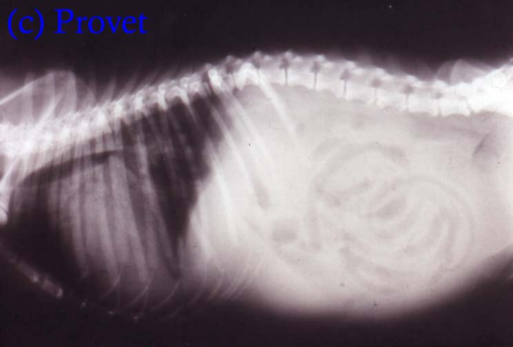
The XRay below is of a terrier showing some of the classical radiographic changes associated with heart failure including :
- Enlarged heart
- Displacement of trachea upwards (dorsally) towards the spine
- Fluid accumulation in the lungs
- Swollen abdomen
- Fluid free in the abdomen (ground glass appearance)
- Enlarged liver
- Poor body musculature (over top of spine)
Treatment
There are 3 main ways in which to manage heart failure patients :
1) Failure of heart muscle -improve cardiac muscle efficiency by the use of positive inotropic drugs (e.g. pimobendan, digoxin), controlling any dysrhythmias - e.g. slow an increased heart rate (tachycardia) improve blood supply to the heart muscle by the use of calcium antagonists that dilate the coronary blood vessels.
2) Excessive circulating blood volume - reduce volume overload and dilation of the heart by the use of diuretics (e.g. frusemide or potassium-sparing diuretics like spironolactone), the use of ACE-inhibitors (enalapril and benazepril) or venodilators (e.g. nitroglycerine) and restriction of salt intake
3) Excessive pressures on the heart - reduce the workload on the heart through rest, by reducing arterial pressure (afterload) and venous pressures (preload) on the heart by the use of vasodilator drugs - benazapril, enalapril, ramipril, imidapril, hydralazine, nitroglycerine. Diuretics can reduce preload through fluid loss from the circulation. Weight loss is important in obese animals
Theophylline is licensed for use in heart failure in the UK. It dilates coronary arteries and increases the strength of contraction of the myocardium. It also acts on the kidney to induce diuresis.
Prognosis
Guarded
Long term problems
Updated November 2013
d>