|
PULMONIC STENOSIS This information is provided by Provet for educational purposes only. You should seek the advice of
your veterinarian if your pet is ill as only he or she can correctly advise
on the diagnosis and recommend the treatment that is most appropriate for
your pet.
Note for Pet Owners:
Topics on this Page: |
Description
Pulmonic stenosis is a congenital narrowing in the region of the pulmonary valve which lies between the right ventricular chamber of the heart and the pulmonary artery. This artery carries deoxygenated blood from the heart to the lungs and the narrowing impairs normal blood flow into the artery. The narrowing can occur within the valve itself (valvular)- commonest site; below the valve at the conus arteriosus or infundibulum (subvalvular) ; or above the valve in the pulmonary artery itself (supravalvular)- rare. Sometimes more than one form is seen in an individual and, although usually an isolated lesion, pulmonic stenosis can occur with other defects and it is one of the components of another congenital defect Tetralogy of Fallot.
History and presenting signs
Usually, affected animals show no external signs of the disease and it is frequently diagnosed during routine examination of the heart with a stethoscope (auscultation). However if blood flow is seriously impaired signs associated with right-sided heart failure may be seen including poor exercise tolerance and feinting (syncope). Sudden death is also sometimes possible.
Physical examination
An abnormal heart sound (called a murmur) may be heard when listening with a stethoscope (called auscultation). The murmur occurs during the systolic phase of the cardiac cycle - when the ventricles are contracting forcing blood out of their chambers and into the pulmonary artery (from the right ventricle) or aorta (from the left atrium).
Electrocardiography (ECG)
On ECG examination there are periods of increased rate of electrical discharges with an abnormal rhythm - called tachydysrhythmia. These result from poor blood supply (ischaemia) to the heart muscle, and so lack of oxygen, which damages the heart muscle resulting in abnormal (ectopic) electrical discharges. These tachydysrhythmias can lead to feinting or death.
XRay
Enlargement of the right ventricle may be seen and, in some cases, bulging of the pulmonary artery can be seen on a dorsoventral view.
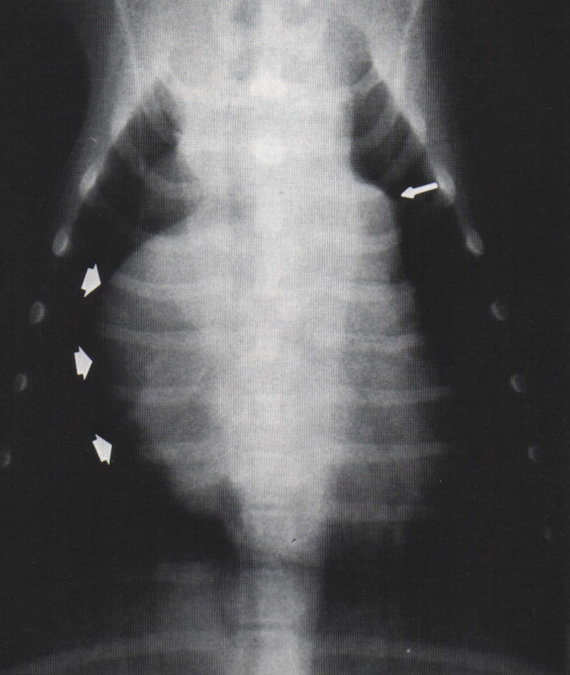
Dilatation of the pulmonary artery (thin arrow at 2 o'clock position) and ventricular enlargement in a dog with pulmonic stenosis. This photograph is reproduced from "Canine & Feline Cardiology" by Phillip R Fox with the permission of Elsevier.
Enlargement of the right atrium may be seen on the dorso-ventral view in animals with tricuspid valve regurgitation.
The special imaging technique by which contrast media is passed through the heart chamber and the sequential X-rays are taken (angiography) is very helpful to confirm the diagnosis.
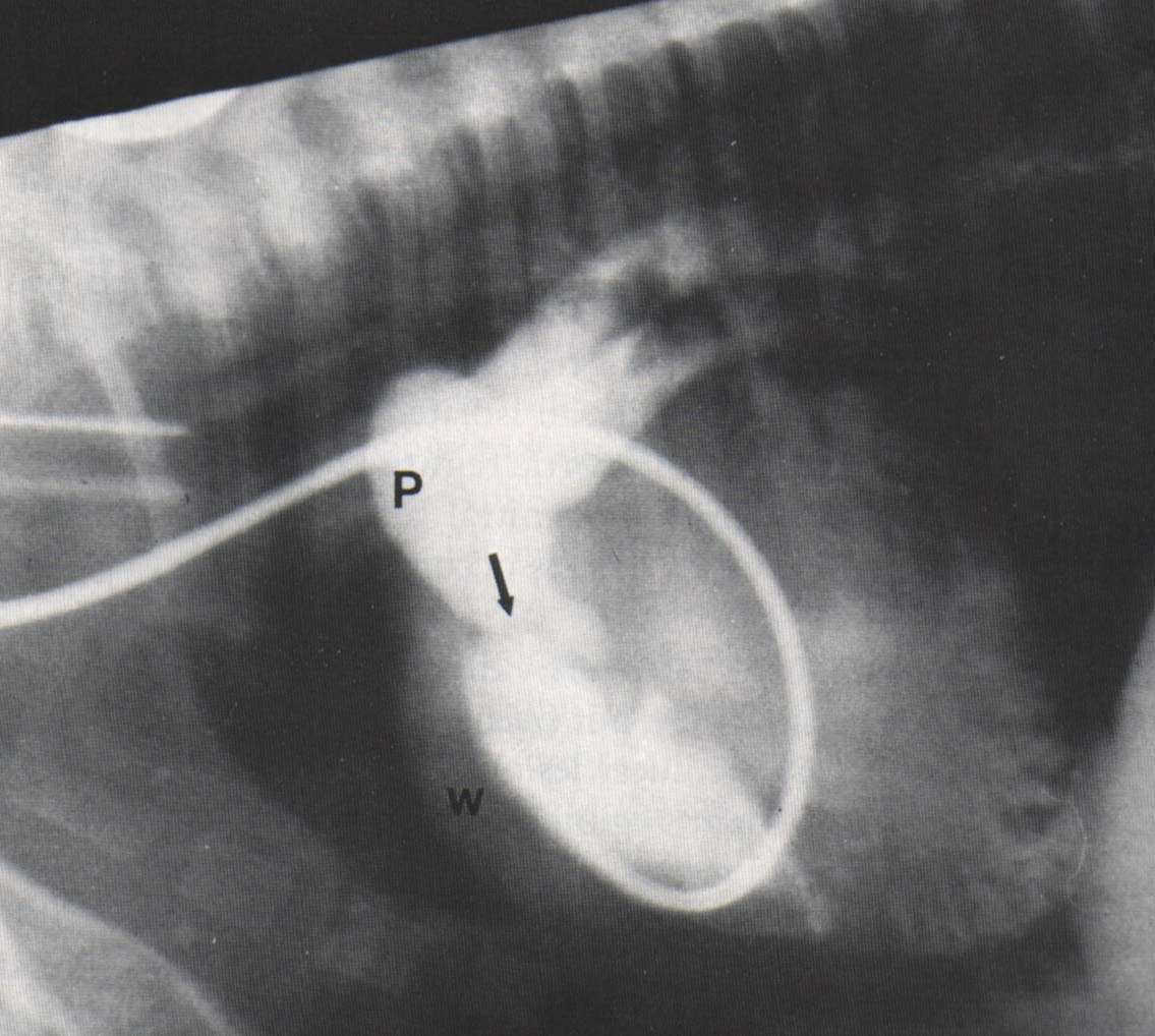
Angiography showing dilatation of the pulmonary artery (P), thickening of the wall of the right ventricle due to hypertrophy (W) and thickening and dysplasia of the pulmonic valve (arrow) in a dog with valvular pulmonic stenosis. This photograph is reproduced from "Canine & Feline Cardiology" by Phillip R Fox, with the permission of Elsevier .
Species Variations
DogsChihuahuas, English Bulldogs and Fox Terriers as having a higher prevalence for the disease than the general population (Patterson Dog. Circ.Res 23:171, 1968). Another report (Canine & Feline Cardiology by Phillip R Fox 1988) also cites Beagles, Samoyeds and Cocker Spaniels as having increased risk. In a recent UK publication (Diseases of the Cardiorespiratory System by Mike Martin and Brendon Corcoran, 1997) the Miniature Schnauzer and Boxer are also listed as being commonly affected, but Samoyeds, Chihuahuas, Fox Terriers and English Bulldogs are not mentioned. Pulmonic stenosis has been shown to be hereditary in Beagles and is thought to be polygenic (not a single gene expression, but multiple genes are involved). In Breed Predispositions to Disease in Dogs and Cats by Gough and Thomas the Mastiff is mentioned.
In dogs pulmonic stenosis is one of the most frequently recognised congenital cardiac abnormalities in dogs estimated to occur in 1/1000 puppies. The literature is confusing regard to breeds most often affected. One study cited
Treatment
Most animals with pulmonic stenosis do not require treatment. Those showing clinical signs of heart failure are treated with diuretics(e.g. frusemide).
Severely affected cases can be treated using a relatively new technique called balloon valvuloplasty in which a catheter with a small balloon on the end is guided to the position of the narrowing, and the balloon is then inflated to open the constriction. This procedure appears to be good in the short-term, but long-term studies are needed.
Updated January 2016
d>