
|
HYPERTROPHIC OSTEODYSTROPHY (HOD) This information is provided by Provet for educational purposes only. You should seek the advice of your veterinarian if your pet is ill as only he or she can correctly advise on the diagnosis and recommend the treatment that is most appropriate for your pet. Topics on this Page: |
Description
Hypertrophic osteodystrophy is an extremely distressing condition which affects young rapidly growing breeds of dog. The disease causes acute pain, debilitation and death in some cases. The disease affects the metaphyseal region of the long bones, and, although any bone can be involved, the bones most often affected are those of the lower limbs (ie below the elbow in the foreleg, and below the stifle in the hindleg).
Cause
The cause of hypertrophic osteodystrophy is unknown.
Early reports that some dogs responded to vitamin C administration, despite the fact that dogs can manufacture their own vitamin C and do not have a dietary requirement for it. This lead to speculation that deranged vitamin C metabolism may be involved in the disease. However, subsequent reports suggest that vitamin C supplementation may be contraindicated because it increases serum calcium concentrations.
The observation of soft tissue calcification in some dogs suggest that mineral imbalance, overnutrition or abnormal hormonal control of calcium and/or phosphorus metabolism may be involved (e.g. mediated by vitamin D, or parathyroid hormone).
Breed Occurence
Hypertrophic osteodystrophy affects young, rapidly growing dogs and the following breeds have been reported to be affected :
Age
Most animals are affected between 2-8 months of age.
Sex
Studies suggest that males may be slightly more affected than females.
Signs
Signs of the disease can be mild with a slight lameness but they are often extremely severe with hard, painful swellings of the long bones which are warm to the touch. Severely affected dogs have a high body temperature, depression, refuse to eat and are unable to walk. This presents management problems for owners of large or giant breeds of dog.
Periods of remission can occur followed by relapse. Death can occur and euthanasia is often requested by owners with severely affected individuals. Puppies that survive this disease may have permanent skeletal deformities such as cow-hocks or angular deformity of the front legs.
Diagnosis
Xrays are needed to identify the characteristic changes of a radiolucent (black) line above the epiphysis and new bone laid down around the long bones. This new bone can be deposited either under or outside the periosteum and it has a rough, irregular appearance and is not smooth as in other disorders (e.g.
The Xray below shows the roughened outline of the long bones due to irregular new bone deposition. There is also soft tissue swelling.
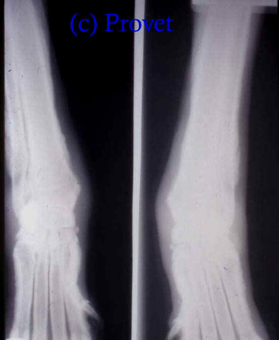
In some cases soft tissue calcification may be present as well (e.g. the aorta, lining of the heart (endocardium), kidneys or lungs.
Treatment
Many therapeutic agents have been used without success. Pain relief is needed for severely affected animals, and nutritional support is needed if they are unable to eat voluntarily. Parenteral fluid administration may be needed in dehydrated animals. Dietary imbalances should be corrected if they are present. Over supplementation with vitamins or minerals should be avoided.
Prognosis
Favourable except in the most severely affected cases.
Relapses are rare once skeltal maturity is reached.
Updated October 2013
d>