
|
MALASSEZIA DERMATITIS Note for Pet Owners:
This information is provided by
Provet for educational purposes only.
You should seek the advice of
your veterinarian if your pet is ill as only he or she can correctly advise
on the diagnosis and recommend the treatment that is most appropriate for
your pet. Topics on this Page:
|
Description
Malassezia dermatitis is a secondary inflammatory skin disease caused by the
presence of a yeast - Malassezia pachydermatis in dogs, Malassezia
sympodialis in cats.
Cause
Malassezia sympodialis has been reported to cause dermatitis in cats
Breed Occurrence
The following breeds of dog are reported to have an increased risk of developing
Malassezia dermatitis : the Australian Terrier, Bassett Hound, Chihuahua,
Dachshund, German Shepherd Dog, Maltese, Newfoundland, Poodle, Silky, Shetland
Sheepdog, Springer Spaniel, and the West Highland White
Terrier
Malassezia dermatitis has also been reported to occur in cats
Signs
Th
e typical clinical signs associated with Malassezia dermatitis in dogs are :- Alopecia
- Erythema
- Greasy coat
- Hyperpigmentation
- Increased scale production
- Lichenification
- Pruritus
- Seborrhoeic smell
Distribution of the lesions may be :
- Focal
- Multifocal
- Generalised
and they most often affect the :
- Abdomen - ventral
- Axillae
- Ear pinnae
- Face
- Feet
- Front legs
- Skin folds
In cats the typical signs are :
- Acne
- Generalised exfoliation
Complications
Diagnosis
Skin cytology is recommended to confirm the diagnosis and suitable samples may
be collected by a variety of techniques including :
- Direct impression smears onto glass slides
- Swabbing
- Sticky tape collection
- Skin scraping
- Biopsy
The yeast can be identified under a microscope by it's characteristic appearance :
- No mycelia
- Lipophilic (stains blue with Dif QuikTM)
- An elongated oval (peanut) shape
- A thick wall
- Budding at one end
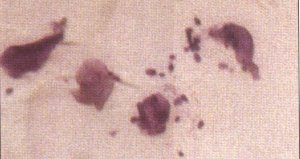
Picture reproduced with permission of the Publishers from Skin Diseases of the Dog and Cat by Harvey and McKeever - available from Provet's On-line Discount Bookshop Click Here
There are circumstances when it is likely to have an important
role to play in disease, notably in association with the following conditions
:
Treatment
Any primary underlying disease must be treated. In addition, specific therapy
can be given to treat the Malassezia : Anti-inflammatory drugs such as corticosteroids may not provide relief for
the pruritus seen in patients with Malassezia dermatitis Prognosis Long term problems Recurrent infections if the underlying problem is not treated successfully Updated October 2013
Good providing an underlying cause can be identified and treated